A stone is a hard, solid mass that can form in the gallbladder, bladder, and kidneys. These types of stones have different causes and are treated in different ways. This leaflet discusses kidney and ureteral stones. These develop in the kidney and either stay there or move to the ureter. Kidney stones form when minerals or acid salts in your urine crystalize. Most stones leave your body while you urinate. However, in some cases you may need treatment to remove the stone. Anyone may develop a kidney stone during his or her lifetime. Stones can form if there is an imbalance in the way your body produces urine. This may be connected to how much you drink and whether there are substances in your urine which trigger stone formation.
People often associate kidney and ureteral stones with pain. However, symptoms can vary from severe pain to no pain at all, depending on stone characteristics – such as the size, shape, and location of the stone in the urinary tract.
Not all stones require treatment. You need treatment if your stone causes discomfort and does not pass naturally with urine. Your doctor may also advise treatment if you have pre-existing medical conditions.
If you have a kidney or ureteral stone which does not cause discomfort, you will generally not receive treatment. Your doctor will give you a time schedule for regular control visits to make sure your condition does not get worse. If your stone is likely to pass with urine, your doctor can prescribe drugs to ease this process. This is called conservative treatment.
Conservative stone treatment
Most kidney or ureteral stones will leave your body while you urinate. However, depending on the size and location of the stone, it will take you some time to pass the stone. You may suffer from renal colic when the stone moves.
In general you can keep this in mind:
• The closer the stone is to the bladder, the higher the chance of passing it
• The bigger the stone, the smaller the chance of passing it
Medical Expulsive Therapy
Your doctor may prescribe drugs (so called alphablockers ,or nifedipine) to help you pass the stone faster and to limit pain while it moves. This is called
Medical Expulsive Therapy (MET) and it is most effective for ureteral stones. During MET you should see your doctor regularly – how often depends on his or her recommendation. The doctor needs to check if the stone keeps moving and if your kidneys continue to function well.
Symptoms
Dull pain or no symptoms at all
Stones can also cause a recurrent, dull pain in the flank. This kind of pain may be a symptom of other diseases as well, so you will need to take medical tests to find out if you have kidney or ureteral stones. Some stones do not cause any discomfort. These are called asymptomatic stones and are usually small. In general asymptomatic stones are found during x-ray or similar imaging procedures for other conditions.
Anyone may develop a kidney stone during his or her lifetime. Stones can form if there is an imbalance in the way your body produces urine. This may be connected to how much you drink and whether there are substances in your urine which trigger stone formation.
SWL is done with a machine that can break stones from outside the body. To break the stone, focused shock waves (short pulses of high energy sound waves) are transmitted to the stone through the skin. The stone absorbs the energy of the shock waves and this breaks it into smaller pieces. The stone fragments then pass with urine in the days or weeks after the procedure
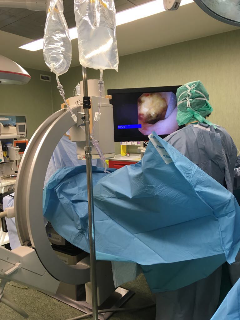
PNL is a surgery to remove large stones directly from the kidney. The advantage is that even very large stones are removed in a single operation. PNL is carried out under general anaesthesia.
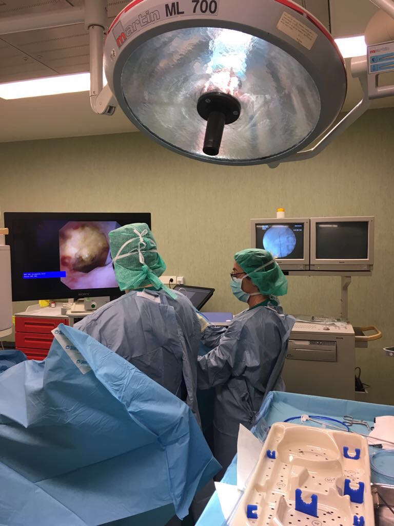
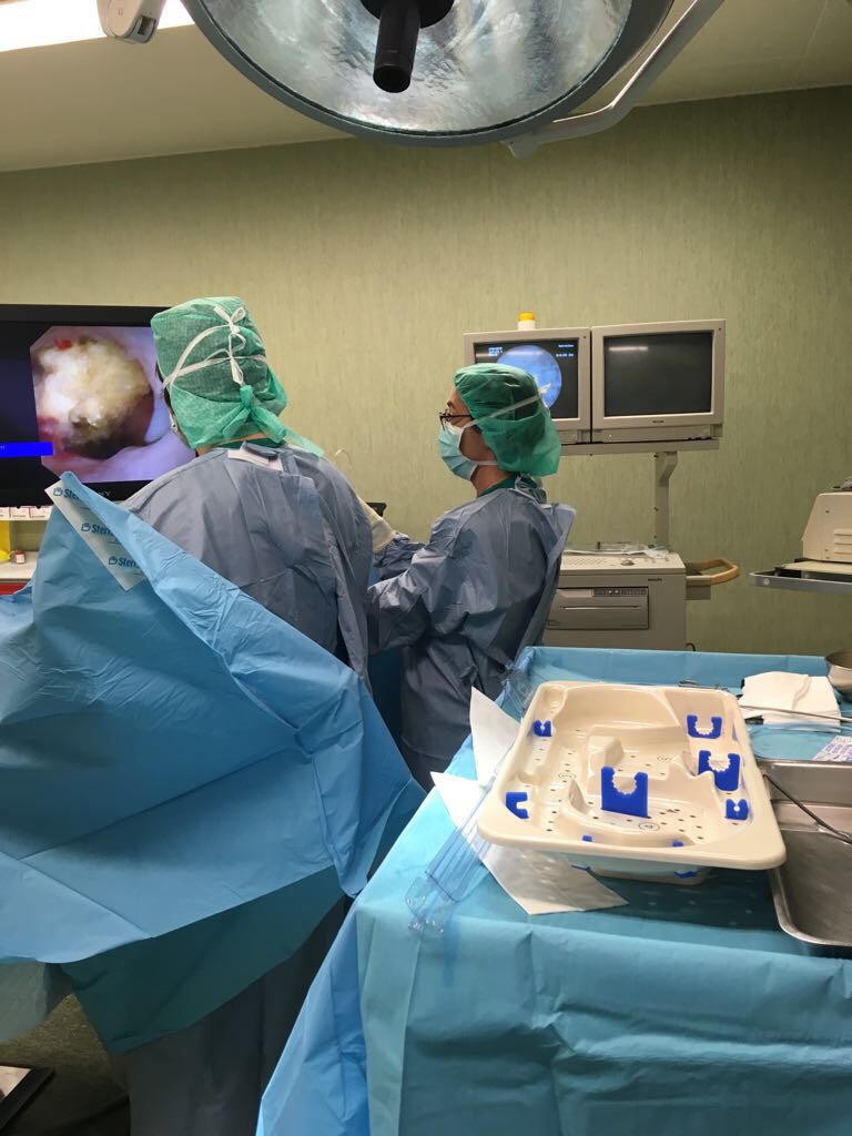
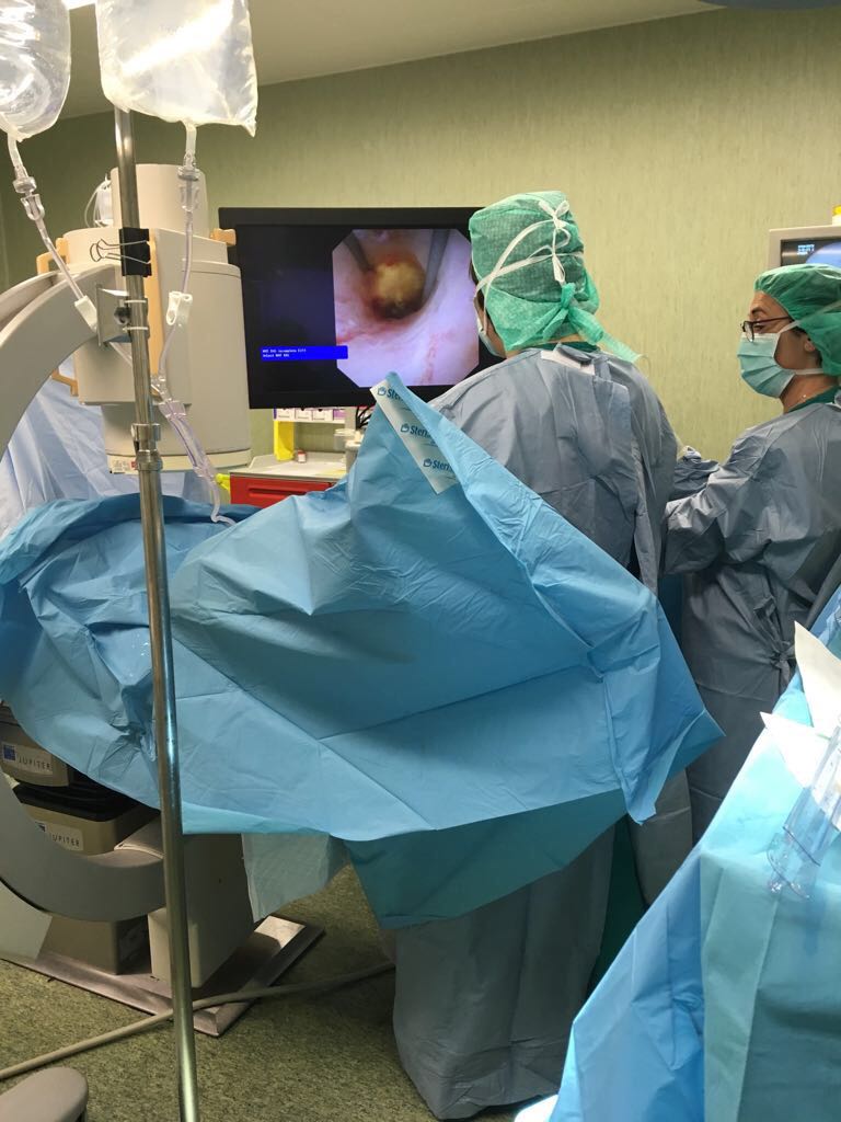
URS is a type of treatment which is done with a small-calibre endoscope. URS is common, success rates are very high, and the risk of complications is low.
For URS you will receive general or local anaesthesia. Once you are under anaesthesia, the doctor enters your bladder with the endoscope through the urethra without making an incision in your body. The stone is pulled out using a special “basket .
The doctor does a series of tests to understand what causes your symptoms. This is called a diagnosis. First, the doctor or nurse will take your medical history and do a physical examination. Then, they will take images of your body and perform other tests if needed.
Imaging techniques
To locate your stone the doctor needs to take images of your internal organs. You will get an ultrasonography (also known as ultrasound), which uses high-frequency sounds to create an image. In addition to ultrasonography, you may need an x-ray of the urinary tract.
Another common method of diagnosis is a CT-scan (computed tomography). This scan can clearly show the size, shape, and thickness of the stone.
Stone analysis and other tests
In case of renal colic, your urine and blood is tested to see if you have an infection or kidney failure. If your stone is expected to pass with urine, your doctor may recommend that you filter your own urine to collect the stone. The doctor will analyse it in order to understand what type of stone you have. This information is important because it helps to select the best options for treatment and prevention.
Your doctor may prescribe drugs (so called alphablockers or nifedipine) to help you pass the stone faster and to limit pain while it moves. This is called Medical Expulsive Therapy (MET) and it is most effective for ureteral stones.
During MET you should see your doctor regularly – how often depends on his or her recommendation.
The doctor needs to check if the stone keeps moving and if your kidneys continue to function well.
Kidney or ureteral stones should be treated if they cause symptoms. There are three common ways to remove stones: shock-wave lithotripsy (SWL), ureteroscopy (URS), and percutaneous nephrolithotomy (PNL). Which active treatment option is best for you depends on many aspects. The most important factor is the symptoms the stone causes. Based on whether the stone is in your kidney or your ureter, the doctor may recommend different treatment options.
Drink more
• Make sure you drink 2.5 to 3 litres every day
• Drink evenly throughout the day
• Choose pH-neutral drinks such as water or milk
• Monitor how much you urinate. It should be 2 to 2.5 litres every day
• Monitor the colour of your urine: it should be light
• Drink even more if you live in a hot climate or do a lot of physical exercise. This will help you to balance your fluid loss
Adapt your diet
Depending on your individual situation, your doctor may recommend that you adapt your diet. It is important to discuss this with the doctor first.
• Have a balanced and varied diet
• Eat lots of vegetables, fibres, and fruits (especially
citrus fruits)
• Try to eat more low-oxalate foods like eggs, lentils, white rice, peeled apples, grapes, cauliflower, squash, etc.
• Make sure your diet contains a sufficient amount of calcium (about 1,000 milligrams a day).
However be careful with calcium supplements and always ask your doctor or nurse for advice
• Reduce the amount of salt in your diet (no more than 3 to 5 grams a day)
• Do not eat too much animal protein, especially meat from young animals. Instead, eat more vegetable protein, found for example in avocados, cauliflower, or peas
• Maintain a healthy weight (your Body Mass Index should be between 18-25 kg/m2)
Healthy habits
Adopting a healthy lifestyle is always a good idea.
• Try to exercise 2 or 3 times a week
• Avoid stress
Some patients who have had kidney or ureteral stones may form more stones in the future. After your stone passes or is removed, your doctor will determine if you are at high risk of recurrence. To do so, he or she will need to analyse the stone. In addition, the doctor will consult the results of your blood and urine tests which were done before treatment. If your risk of recurrence is low, general lifestyle changes will be enough to cut the risk of forming another stone.
The form of these stones is similar to that of some deer antlers as they grow to fill the inside of the rim. Generally, the coraliforme lithiasis is formed due to repeated urinary tract infections (UTI) with certain types of bacteria. Although they can grow very large, you may not know you have them completely, since they cause little or no pain.
Usually not. Commonly, we heard a patient complaining of pain lower back kidney pain but most often is not in fact what happens. Usually these pain are muscle-skeletal and do not have exactly to do with the kidney pain.
Pain of renal origin, caused by infection or for another reason as for example stones, is a pain in the lower back, which usually has no relief position (antiallergic position) and often radiates to the anterior region, and may even refer to irradiation to the groin area (testis in man and vagina in woman). This pain may still be accompanied by fever, nausea and/or vomiting.
During a renal colic episode, the symptoms usually include severe sudden onset pain, in which the patient denies any antiallergic position. The pain usually starts suddenly causing irritation and obstruction. Typically, the pain has the ipsilateral lumbar location with sometimes anterior irradiation to the groin or abdomen. The appearance of blood in the urine, nausea and / or vomiting is also common.
Not necessarily. The patient can develop stones, and even reach an important size, which will only feel symptoms, if these stones partially block the passage of urine, and thus trigger pain. This pain, this colic is also related to the shape, size and location of the stone.
The stones are formed when there is an imbalance between certain urinary chemical components, such as calcium, oxalate and phosphate. Some of these chemical components promote crystallization, while others inhibit it. A less common type of stone is caused by an infection in the urinary, this type of stone is called struvite or stone of infection. Less common are the pure stones of uric acid. Much rarer is the case of the hereditary type of cystine stones and even rarer are those linked to other hereditary diseases.
For a number of reasons, the number of patients with kidney stones has been increase over the last 20 years. Although the number of patients with stones are mostly male, the number of women has also increasing. Climate, low water intake, salt-rich diets an important role in the formation of stones in the urinary system and later on the appearance of renal colic. There are also certain foods that can promote the formation of stones in more susceptible people. There are also factors such as family or personal history, such as certain diseases, urinary tract infections, repetition, which may constitute a risk factor for the formation of stones.
Yes this is true. Of course there are certain aspects such as family history, anatomical changes, or a history of early-stage renal colic that is a risk factor for the development of kidney stones. Of course this is why stone development can occur and the patient does not have any symptoms until the first episode of renal colic.
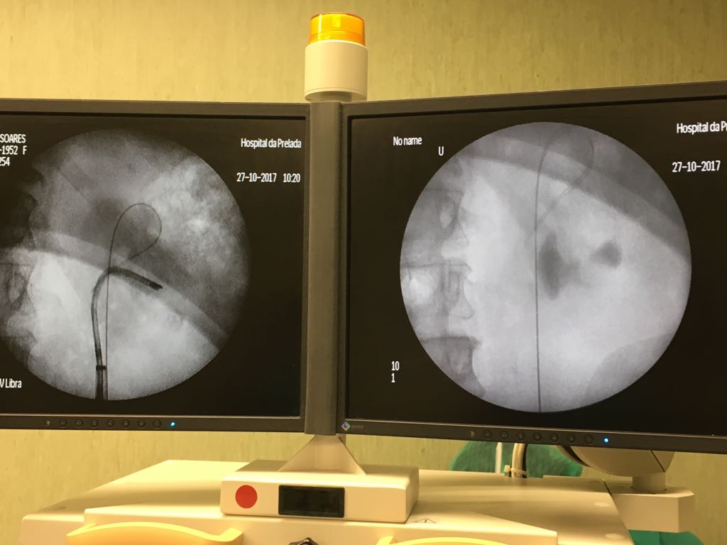
X-ray and ultrasound are tests that give us important information on the existence of stones or not, but there are others the TAC that allows us to not only diagnose and locate with great precision calculations as well as being able to program a possible treatment if it is if any.
We have to differentiate two types of patients, the patient in urgent/acute renal colic and one in which is not in an acute situation in which the treatment can be scheduled, in both cases, undergoing surgery or medical treatment.
Here we have the technology next to Urology, that is, the vulgar surgery open to remove stones, practically, except for very rare exceptions currently no longer practiced. Currently through the urethra, bladder, urethra and kidney we reach the stone, or directly through a hole with 2 cm we come directly to the kidney when we are talking about stones in a size greater than 2/3 cm.
The discussion with your Urologist is fundamental for planning the its diagnosis, treatment and adoption of a lifestyle that does not potentiate either the growth of existing stones or the formation of new stones. Adoption of a proper diet, abundant water intake, avoidance of intake of foods rich in oxalate such as leafy vegetables, nuts, tea or chocolate should be taken into account.